With
the introduction of new excitation/detection schemes in microscopy,
such as
confocal, multiphoton and total internal reflection, and the advances
in
fluorophore technology, fluorescence microscopy is today the most
rapidly
growing light microscopy technique, both in medical and biological
sciences.
In-vivo imaging with high lateral and axial resolution and large
penetration
depth can be obtained with confocal microscopy [1]. This technique is
selecting the
signal from a slice of the object around the focal plane by using a
pinhole in
the detection that attenuates out-of-focus light. However, confocal
microscopy
is exciting a large sample volume, and detecting only a small fraction
of it (the focal volume), thus creating significantly more
photoexcitation than needed. This problem can be overcome by using
two-photon
(i.e.
non-linear) fluorescence microscopy in which two photons with a
wavelength in
near-infrared region are absorbed [2]. Significant two-photon
absorption is
reached
only in the focal volume where high photon densities are present,
allowing for an
intrinsic 3D spatial resolution. Moreover, near infrared light is less
scattered than the visible light, with consequent improvement of the
image contrast
and penetration depth.
Besides its many advantages, fluorescence
microscopy has some limitations. Background
fluorescence originates from endogenous object constituents
(autofluorescence)
or from unbound or non-specifically bound fluorescent probes (reagent
background), and fluorophores under light excitation
are prone
to irreversible degradation (photobleaching).
Recently,
colloidal quantum dots (CQDs) have been demonstrated as innovative
fluorophores
because of their unique optical properties
[3]. CQDs
are semiconductors nanocrystals (e.g. CdSe core / ZnS shell) of a few
nanometers in
diameter. The wavelength of the emitted light depends on their size and
shape
that can be controlled during their synthesis (see Fig. 1) [4].
They show
an
increasing absorption towards shorter wavelengths, resulting in a
broad
absorption spectrum, in stark contrast to organic dyes, which feature
an absorption peaked about 50-100nm blueshifted to the emission,
strongly decaying twoards shorter wavelengths. It is therefore much
more practical to excite CQDs
emitting at different wavelength by a single monochromatic light
source, simplifying multiple
marker
imaging (Fig. 2). Furthermore, the emission spectrum of CQDs is
narrower than that of dyes, enabling an improved spectral
discrimination of the emission from multiple CQD markers.
CQDs are also attractive for two-photon fluorescence
microscopy
due to their high two-photon absorption cross-section. CQDs exhibit
high
photostability as compared to organic dyes, however surface defects in
the
crystal structure can act as temporary traps for carriers, preventing
their
radiative recombination. Furthermore, charging of the CQDs can
significantly reduce their quantum yield, The temporal sequence of
charging and trapping events
often results in intermittent fluorescence (photoblinking) [5], a
limitation
for CQDs
application in fluorescence microscopy.
Beyond
fluorescence, we can use CQDs excited in resonance it fundamental
absoprtion peak to create a strong non-linear
signal
called four-wave mixing (FWM) which is due to its third-order
non-linearity [6]. The aim of this project is to study the
applicability of
such resonant
FWM signal from CQDs as biomarkers both in-vitro and in-vivo, to obtain
a novel
type of multi-photon imaging, with intrinsic sectioning capability
without the
need of a detection pinhole to reject out of focus light, alternative
to
two-photon fluorescence microscopy. Since FWM is a coherent signal
directly
linked to the third-order polarization, it has the benefit of detection
free
from any incoherent fluorescence background. Moreover, the spatial
resolution
can be increased beyond the diffraction limit due to the optical
non-linearity.
Project Aims
- Implementation of a set-up to measure and image the transient resonant FWM on biocompatible CQDs in solution with sub-micron three-dimensional spatial resolution.
- Quantitative evaluation of the sensitivity of the technique and the related CQD photostability by investigating the FWM signal intensity and long-term stability versus CQD density and excitation power.
- Determination of the lateral and axial spatial resolution of the technique by imaging of reference structures such as patterned arrays of CQDs in polymer matrices.
- Imaging of cells (both fixed and living) labelled with CQDs to assess the applicability of the technique to cell biology.
- Demonstration of zero-background imaging performance by the implementation of a three-pulse FWM.
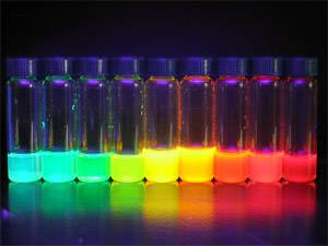 Fig.1:
CdSe/Zns colloidal quantum dots emit different color wavelenghts depending on their size.
 Fig.2:
Absorption and emission spectra of CdSe/Zns colloidal quantum dots with different size.
